Ophthalmoscope Retinal Detachment . Retinal detachment occurs when subretinal fluid accumulates between the neurosensory retina and the retinal pigment epithelium. Symptoms of retinal detachment may include the following: Indirect ophthalmoscopy shows the retinal detachment and can differentiate the subtypes of retinal detachment in nearly all cases. This process can occur in three ways. Retinal detachment is separation of the neurosensory retina from the underlying retinal pigment epithelium. Retinal detachments (rds) constitute a severe ocular condition that can lead to permanent vision loss. Retinal detachment (rd) is an acute or progressive condition in which the neuroretina separates from the retinal pigment. Photopsia (common initially) visual field defect (developing over time; The ophthalmologist may use an instrument with a bright light and a special lens to examine the inside of your eye. Direct funduscopy using a handheld ophthalmoscope can.
from webeye.ophth.uiowa.edu
Symptoms of retinal detachment may include the following: This process can occur in three ways. Retinal detachment is separation of the neurosensory retina from the underlying retinal pigment epithelium. Retinal detachment occurs when subretinal fluid accumulates between the neurosensory retina and the retinal pigment epithelium. Retinal detachments (rds) constitute a severe ocular condition that can lead to permanent vision loss. Direct funduscopy using a handheld ophthalmoscope can. Photopsia (common initially) visual field defect (developing over time; The ophthalmologist may use an instrument with a bright light and a special lens to examine the inside of your eye. Indirect ophthalmoscopy shows the retinal detachment and can differentiate the subtypes of retinal detachment in nearly all cases. Retinal detachment (rd) is an acute or progressive condition in which the neuroretina separates from the retinal pigment.
Atlas Entry Rhegmatogenous retinal detachment
Ophthalmoscope Retinal Detachment Symptoms of retinal detachment may include the following: Direct funduscopy using a handheld ophthalmoscope can. Photopsia (common initially) visual field defect (developing over time; Retinal detachment is separation of the neurosensory retina from the underlying retinal pigment epithelium. Retinal detachments (rds) constitute a severe ocular condition that can lead to permanent vision loss. Indirect ophthalmoscopy shows the retinal detachment and can differentiate the subtypes of retinal detachment in nearly all cases. Retinal detachment (rd) is an acute or progressive condition in which the neuroretina separates from the retinal pigment. Symptoms of retinal detachment may include the following: Retinal detachment occurs when subretinal fluid accumulates between the neurosensory retina and the retinal pigment epithelium. This process can occur in three ways. The ophthalmologist may use an instrument with a bright light and a special lens to examine the inside of your eye.
From geekymedics.com
Retinal Detachment Ophthalmology Geeky Medics Ophthalmoscope Retinal Detachment Retinal detachment is separation of the neurosensory retina from the underlying retinal pigment epithelium. Indirect ophthalmoscopy shows the retinal detachment and can differentiate the subtypes of retinal detachment in nearly all cases. Retinal detachment occurs when subretinal fluid accumulates between the neurosensory retina and the retinal pigment epithelium. Retinal detachment (rd) is an acute or progressive condition in which the. Ophthalmoscope Retinal Detachment.
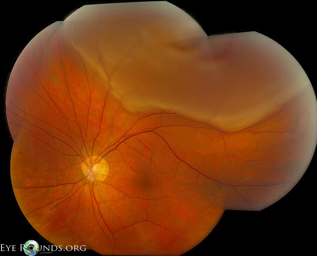
From eyerounds.org
Atlas Entry Rhegmatogenous retinal detachment Ophthalmoscope Retinal Detachment Indirect ophthalmoscopy shows the retinal detachment and can differentiate the subtypes of retinal detachment in nearly all cases. Retinal detachment is separation of the neurosensory retina from the underlying retinal pigment epithelium. Direct funduscopy using a handheld ophthalmoscope can. This process can occur in three ways. Symptoms of retinal detachment may include the following: Photopsia (common initially) visual field defect. Ophthalmoscope Retinal Detachment.
From webeye.ophth.uiowa.edu
Retinal Detachment From One Medical Student to Another Ophthalmoscope Retinal Detachment Photopsia (common initially) visual field defect (developing over time; Indirect ophthalmoscopy shows the retinal detachment and can differentiate the subtypes of retinal detachment in nearly all cases. Retinal detachments (rds) constitute a severe ocular condition that can lead to permanent vision loss. Retinal detachment occurs when subretinal fluid accumulates between the neurosensory retina and the retinal pigment epithelium. Direct funduscopy. Ophthalmoscope Retinal Detachment.
From imagebank.asrs.org
Retinal Detachment Retina Image Bank Ophthalmoscope Retinal Detachment Retinal detachment is separation of the neurosensory retina from the underlying retinal pigment epithelium. Retinal detachment (rd) is an acute or progressive condition in which the neuroretina separates from the retinal pigment. Retinal detachment occurs when subretinal fluid accumulates between the neurosensory retina and the retinal pigment epithelium. The ophthalmologist may use an instrument with a bright light and a. Ophthalmoscope Retinal Detachment.
From rsnallc.com
Complex Retinal Detachment Retina Specialists of North Alabama, LLC Ophthalmoscope Retinal Detachment Direct funduscopy using a handheld ophthalmoscope can. Retinal detachments (rds) constitute a severe ocular condition that can lead to permanent vision loss. Retinal detachment occurs when subretinal fluid accumulates between the neurosensory retina and the retinal pigment epithelium. Photopsia (common initially) visual field defect (developing over time; Indirect ophthalmoscopy shows the retinal detachment and can differentiate the subtypes of retinal. Ophthalmoscope Retinal Detachment.
From www.eyerounds.org
Atlas Entry Rhegmatogenous retinal detachment Ophthalmoscope Retinal Detachment Retinal detachment (rd) is an acute or progressive condition in which the neuroretina separates from the retinal pigment. Direct funduscopy using a handheld ophthalmoscope can. Indirect ophthalmoscopy shows the retinal detachment and can differentiate the subtypes of retinal detachment in nearly all cases. Photopsia (common initially) visual field defect (developing over time; Retinal detachment occurs when subretinal fluid accumulates between. Ophthalmoscope Retinal Detachment.
From www.researchgate.net
Integrated scanning laser ophthalmoscope and ultrawidefield imaging Ophthalmoscope Retinal Detachment This process can occur in three ways. Photopsia (common initially) visual field defect (developing over time; Retinal detachment occurs when subretinal fluid accumulates between the neurosensory retina and the retinal pigment epithelium. Direct funduscopy using a handheld ophthalmoscope can. The ophthalmologist may use an instrument with a bright light and a special lens to examine the inside of your eye.. Ophthalmoscope Retinal Detachment.
From imagebank.asrs.org
Tractional Retinal Detachment in PDR Retina Image Bank Ophthalmoscope Retinal Detachment Retinal detachment occurs when subretinal fluid accumulates between the neurosensory retina and the retinal pigment epithelium. Photopsia (common initially) visual field defect (developing over time; Retinal detachment (rd) is an acute or progressive condition in which the neuroretina separates from the retinal pigment. Indirect ophthalmoscopy shows the retinal detachment and can differentiate the subtypes of retinal detachment in nearly all. Ophthalmoscope Retinal Detachment.
From geekymedics.com
Retinal Detachment Ophthalmology Geeky Medics Ophthalmoscope Retinal Detachment Retinal detachment is separation of the neurosensory retina from the underlying retinal pigment epithelium. This process can occur in three ways. Symptoms of retinal detachment may include the following: The ophthalmologist may use an instrument with a bright light and a special lens to examine the inside of your eye. Retinal detachment (rd) is an acute or progressive condition in. Ophthalmoscope Retinal Detachment.
From retinanevada.com
Detached and Torn Retina Las Vegas Retina Consultants of Nevada Ophthalmoscope Retinal Detachment Retinal detachment occurs when subretinal fluid accumulates between the neurosensory retina and the retinal pigment epithelium. Symptoms of retinal detachment may include the following: Photopsia (common initially) visual field defect (developing over time; Direct funduscopy using a handheld ophthalmoscope can. Retinal detachment is separation of the neurosensory retina from the underlying retinal pigment epithelium. Indirect ophthalmoscopy shows the retinal detachment. Ophthalmoscope Retinal Detachment.
From frontrangeretina.com
Symptoms of a retinal detachment Front Range Retina Ophthalmoscope Retinal Detachment Retinal detachments (rds) constitute a severe ocular condition that can lead to permanent vision loss. Retinal detachment occurs when subretinal fluid accumulates between the neurosensory retina and the retinal pigment epithelium. Indirect ophthalmoscopy shows the retinal detachment and can differentiate the subtypes of retinal detachment in nearly all cases. Retinal detachment is separation of the neurosensory retina from the underlying. Ophthalmoscope Retinal Detachment.
From www.pinterest.ca
FUNDOSCOPY Ophthalmology Optometry school, Eye anatomy, Eye facts Ophthalmoscope Retinal Detachment Photopsia (common initially) visual field defect (developing over time; Retinal detachments (rds) constitute a severe ocular condition that can lead to permanent vision loss. Direct funduscopy using a handheld ophthalmoscope can. Symptoms of retinal detachment may include the following: Retinal detachment is separation of the neurosensory retina from the underlying retinal pigment epithelium. The ophthalmologist may use an instrument with. Ophthalmoscope Retinal Detachment.
From vmrinstitute.com
Retinal Detachment Treatment Orange County Retina Specialists Ophthalmoscope Retinal Detachment Retinal detachment (rd) is an acute or progressive condition in which the neuroretina separates from the retinal pigment. The ophthalmologist may use an instrument with a bright light and a special lens to examine the inside of your eye. Symptoms of retinal detachment may include the following: Direct funduscopy using a handheld ophthalmoscope can. Retinal detachments (rds) constitute a severe. Ophthalmoscope Retinal Detachment.
From ophabstract.blogspot.com
Retinal Detachment Ophthalmoscope Retinal Detachment Retinal detachments (rds) constitute a severe ocular condition that can lead to permanent vision loss. Retinal detachment (rd) is an acute or progressive condition in which the neuroretina separates from the retinal pigment. Indirect ophthalmoscopy shows the retinal detachment and can differentiate the subtypes of retinal detachment in nearly all cases. Photopsia (common initially) visual field defect (developing over time;. Ophthalmoscope Retinal Detachment.
From www.slideserve.com
PPT Ocular Emergencies PowerPoint Presentation, free download ID Ophthalmoscope Retinal Detachment The ophthalmologist may use an instrument with a bright light and a special lens to examine the inside of your eye. Photopsia (common initially) visual field defect (developing over time; Indirect ophthalmoscopy shows the retinal detachment and can differentiate the subtypes of retinal detachment in nearly all cases. This process can occur in three ways. Symptoms of retinal detachment may. Ophthalmoscope Retinal Detachment.
From www.sciencephoto.com
Ophthalmoscopy of detached retina in AIDS patient Stock Image M112 Ophthalmoscope Retinal Detachment Photopsia (common initially) visual field defect (developing over time; Retinal detachment is separation of the neurosensory retina from the underlying retinal pigment epithelium. Indirect ophthalmoscopy shows the retinal detachment and can differentiate the subtypes of retinal detachment in nearly all cases. The ophthalmologist may use an instrument with a bright light and a special lens to examine the inside of. Ophthalmoscope Retinal Detachment.
From www.pinterest.com
Pin on Holistic Health, Nature's Remedies Ophthalmoscope Retinal Detachment Retinal detachment (rd) is an acute or progressive condition in which the neuroretina separates from the retinal pigment. Retinal detachment occurs when subretinal fluid accumulates between the neurosensory retina and the retinal pigment epithelium. Retinal detachments (rds) constitute a severe ocular condition that can lead to permanent vision loss. Retinal detachment is separation of the neurosensory retina from the underlying. Ophthalmoscope Retinal Detachment.
From webeye.ophth.uiowa.edu
Atlas Entry Rhegmatogenous retinal detachment Ophthalmoscope Retinal Detachment Symptoms of retinal detachment may include the following: Retinal detachment (rd) is an acute or progressive condition in which the neuroretina separates from the retinal pigment. Retinal detachment is separation of the neurosensory retina from the underlying retinal pigment epithelium. Retinal detachments (rds) constitute a severe ocular condition that can lead to permanent vision loss. This process can occur in. Ophthalmoscope Retinal Detachment.